Three young patients were alive without disease years after receiving experimental T cells. The phase 1 trial cannot show what caused those outcomes
More than 31.8 months. More than 41.2 months. More than 51.6 months.
Those are three disease-free intervals documented from the first infusion of an experimental T cell treatment. The first belonged to a 14-year-old whose medulloblastoma had returned multiple times; observation ended when the patient left the study. The second belonged to an 8-year-old with a rare astroblastoma and was still ongoing at the paper’s cutoff. The third belonged to a 14-year-old whose glioblastoma had returned and was also ongoing.
A month count can sound clinical and remote. Here it is the only honest way to hold the human fact in view. The paper does not tell us what those years felt like. It does tell us that three young patients with high-risk brain tumors were alive without documented disease for periods measured in years after receiving the cells.
It does not tell us that the cells caused those outcomes.
That distinction is the spine of this story. ReMIND was open-label, meaning families and clinicians knew which treatment was given. It was also non-randomized: there was no assigned comparison group. And it was phase 1, an early study built first to test whether multi-target T cells could be made, given and studied at a tolerable dose—not whether they improved survival. It recorded three exceptional clinical histories. It also recorded no scan with enough shrinkage or disappearance to meet the trial’s predefined response threshold in its aggressive brainstem-tumor group, two possibly treatment-related serious events involving tumor swelling, and one fatal event serious enough to count against increasing the dose under the trial’s rules.
The years are real. The attribution is uncertain. Both truths deserve the main story.
A treatment made from each patient’s own blood
The treatment is called TAA-T therapy, short for tumor-associated-antigen-specific T cells. T cells are immune cells that can recognize particular molecular fragments. Researchers collected blood from each participant, then grew that person’s own T cells in the laboratory while exposing them to fragments from three tumor-associated proteins: WT1 (Wilms tumor 1), PRAME (preferentially expressed antigen in melanoma) and survivin, a protein involved in cell survival and division. These proteins can be present in pediatric brain tumors and can act as signposts for immune cells, but they are not unique to cancer.
These were not CAR-T cells, which are T cells genetically modified to carry a laboratory-designed receptor—a new sensor for a chosen target. ReMIND used a different approach: it selected and expanded naturally occurring T cells that reacted to the three protein-fragment targets, without genetically engineering them. The cells were tested, frozen and later delivered into a vein. By aiming at three targets rather than one, the researchers hoped to reduce the chance that a tumor made of different kinds of cells could escape because only some cells carried any single target.
How is this different from CAR-T?
CAR-T manufacturing gives T cells a new, artificial receptor so they can recognize a chosen marker directly. TAA-T manufacturing does not add a receptor. It grows a mixed population of unmodified T cells whose existing receptors respond to protein fragments associated with WT1, PRAME or survivin. These are different ways of making an immune-cell treatment; evidence that one approach works in a particular cancer cannot be transferred to the other.
That is a plausible strategy, not a demonstrated clinical mechanism. The trial did not show that the infused cells entered a particular tumor, persisted there or killed it. Its primary questions were more basic: could a usable product be made, could it be administered, what dose should move forward, and what toxicities appeared?
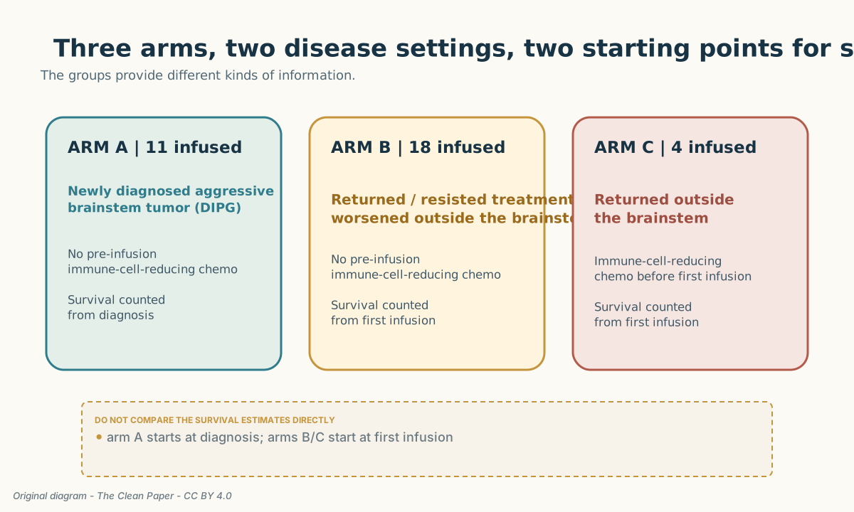
ReMIND enrolled three groups at Children’s National Hospital. Arm A included newly diagnosed diffuse intrinsic pontine glioma (DIPG), an aggressive tumor that grows through the pons, part of the brainstem, and is difficult to remove surgically. Arm B included brain and spinal-cord tumors outside the brainstem that had returned, resisted treatment or worsened. Neither group received lymphodepletion, a short course of chemotherapy that temporarily lowers some of the body’s existing immune cells before an immune-cell infusion. Arm C was a four-patient expansion in non-brainstem tumors that had returned and added this pre-infusion chemotherapy, using fludarabine and cyclophosphamide.
Fifty-one consented; thirty-three were infused
The number of patients narrowed at every practical step.
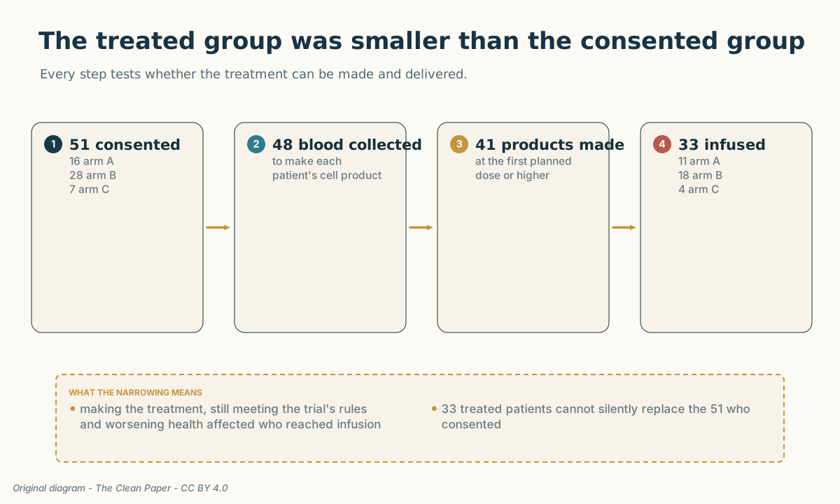
Fifty-one patients consented. Forty-eight had blood collected. Forty-one of those 48, or 85.4%, had a TAA-T product manufactured at the trial’s first planned dose or higher. Thirty-three received at least one infusion: 11 in arm A, 18 in arm B and 4 in arm C.
Some patients became ineligible or died before treatment. Seven patients whose blood had been collected did not reach a usable product at the trial’s first planned dose level: six because the cells did not multiply enough in the laboratory and one because the product failed a required quality check. Nine patients whose first yield was inadequate had to be reassigned to a lower dose.
The process improved during the study. After larger blood collections and changes in how the cells were grown, all 14 patients whose blood was collected after those changes produced at least one treatment dose at the trial’s first planned dose or higher. That is genuine evidence about the ability to make the treatment. It is not a patient-outcome result.
Using its statistical dose-safety model, the study selected a target of 80 million cells per dose for each square metre of estimated body surface area (8 × 10^7 cells/m²). Body surface area is a clinical estimate of overall body size calculated from height and weight; it does not mean that doctors measured a patch of skin, the brain or the tumor. Oncology and pediatric trials use it to scale a planned dose across patients of different sizes—for example, a patient with an estimated body surface area of 1.2 m² would have a target dose of 96 million cells for one infusion. The model also labeled the 80-million-cells-per-m² dose the estimated maximum tolerated dose—the highest dose expected to stay below the trial’s unacceptable-toxicity threshold. The wording matters: the patients actually treated never reached an observed toxicity ceiling. This was a model-based recommendation for a phase 2 study, not proof that the dose is broadly safe.
Most recorded side effects were mild or moderate. One fatal event met the dose-limiting-toxicity rule
An adverse event is any health problem recorded during the study; it is not automatically caused by the treatment. Grades 1 and 2 mean mild or moderate, grade 3 is severe, grade 4 is life-threatening and grade 5 is death. Across ReMIND, 293 of 332 recorded adverse events, or 88%, were grade 1 or 2. Common events included headache, fatigue or lethargy, nausea and vomiting. Three of the 33 infused patients had grade 3 or higher events that appeared or worsened after treatment and were considered at least possibly related to TAA-T therapy. Two patients had possibly related serious events involving edema—swelling in or around the brain or tumor.
One of them was P27, a patient with DIPG. Ten days after infusion, P27 developed hydrocephalus, a dangerous buildup of fluid inside the brain, together with tumor swelling and breathing distress. The patient did not improve after doctors placed a drainage tube called a shunt and increased steroid treatment to reduce swelling. The fatal event was classified as a grade 5 dose-limiting toxicity: under the protocol, it was serious enough to count against further dose escalation.
The trial’s own safety record preserves two attribution judgments at once: the event was considered possibly related to TAA-T therapy and probably related to the underlying disease. Imaging was consistent with tumor progression. The study cannot choose a single cause, and neither should an article about it.
The death prompted a pause in enrollment. The protocol was changed to require a longer period of neurological stability and a shorter interval between radiation and the first infusion. Five more arm A patients were then treated at the same or higher planned doses without another event that met the dose-limiting rule.
The second serious event occurred in P42, who had a worsening diffuse midline glioma in the thalamus, a deep part of the brain. Sixteen days after infusion, magnetic resonance imaging (MRI) showed brain swelling accompanied by headache and other neurologic symptoms. The symptoms returned to their previous level after bevacizumab, a drug that can reduce tumor-associated swelling. This event did not meet the dose-limiting rule.
All four patients in arm C developed temporary grade 3 cytopenias—low blood-cell counts—expected from the pre-infusion chemotherapy; those events were excluded from the combined TAA-T safety analysis. One participant across the study retrospectively met the criteria for mild cytokine release syndrome, a body-wide inflammatory reaction that can occur when immune cells are activated.
The calibrated conclusion is that TAA-T treatment was generally tolerable in this small phase 1 cohort. The unrestricted word safe would erase the size of the study, the two serious swelling events and the fatal dose-limiting toxicity.
What happened across the cohorts
The two clinical populations need to stay separate.
In arm A, the DIPG group, median progression-free survival—the time until the tumor worsened or the patient died—was 10.5 months from diagnosis. Median overall survival—the time patients remained alive—was 13.7 months from diagnosis. Ten patients had scans that could be judged using the protocol’s predefined response rules: six had stable disease, meaning neither enough shrinkage to count as a response nor enough growth to count as progression, and four had progressive disease. No scan showed enough shrinkage or disappearance to meet the trial’s predefined threshold for an objective response.
Arms B and C pooled several non-brainstem tumors that had returned, resisted treatment or worsened because arm C was too small for meaningful comparison on its own. Here, survival was counted from the first infusion, not diagnosis. The median time before tumor worsening or death was 5.0 months, and median time alive was 12.7 months, both measured from infusion. Those estimates cannot be compared directly with arm A’s diagnosis-based numbers.
Among ten patients in arms B and C with measurable disease—at least one tumor area that could be sized reliably on scans—five had stable disease and five had progressive disease. One of the five patients classified as having stable disease, P37, had more than 90% lesion reduction but deteriorated before a second scan could confirm it. A partial response required enough shrinkage to cross the protocol’s predefined threshold and a later scan to confirm it, so the protocol did not classify P37’s result as a partial response.
Five further patients had disease that was nonmeasurable but still evaluable: doctors could judge change on the scans, but no tumor area qualified for dependable size measurement. Their best responses were one complete response, three stable disease and one progressive disease. The complete response belonged to P41. Under the protocol’s scan rules, “complete response” meant that the visible non-target disease disappeared; it did not mean proof of cure. It was not a reliable percentage reduction in a measurable target lesion and does not belong in the ten-patient measurable-disease count.
How do radiologists classify a response?
Before treatment, radiologists identify “target” lesions that are large and clear enough to measure repeatedly. Other visible disease can be followed as “non-target” disease without assigning it a dependable percentage change. A partial response requires enough shrinkage in measured target lesions and usually a later scan to confirm it. A complete response requires disappearance under the applicable scan criteria. These labels describe images at particular times; they do not by themselves prove a cure or identify which treatment caused the change.
Three lives, three different evidentiary stories
The three long intervals are easiest to misread when compressed into one sentence. Their treatment histories and measurement limits were not the same.
P41: a complete response with a difficult baseline
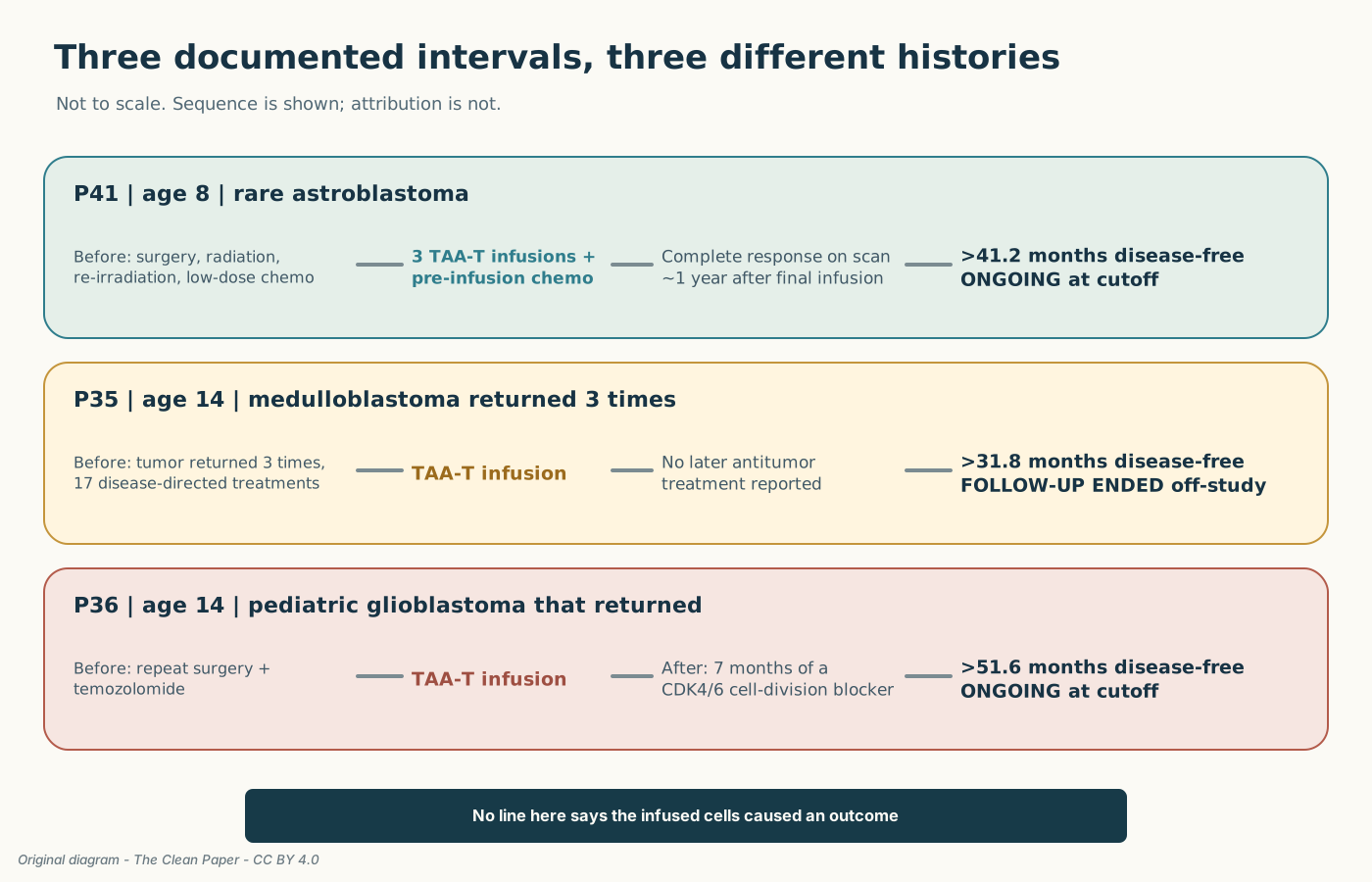
P41 entered the trial at age 8 with an astroblastoma that had returned and carried an EWSR1-BEND2 gene rearrangement, a rare tumor feature in which two genes had joined. It involved the third ventricle, one of the fluid-filled spaces deep in the brain. The child had undergone surgical removal and focused radiation, then a second course of radiation about seven months before TAA-T and low-dose chemotherapy completed about two months before infusion. P41 entered arm C, received the pre-infusion immune-lowering chemotherapy and then three TAA-T infusions.
After specialists re-read the brain scans, the baseline disease was classified as nonmeasurable but evaluable: visible enough to judge, but not suitable for reliable size measurement. About one year after the final infusion, disappearance of the non-target lesion supported the authors’ complete-response classification on imaging. The source data show a disease-free interval of more than 41.2 months from the first infusion, ongoing at the 1 January 2026 cutoff.
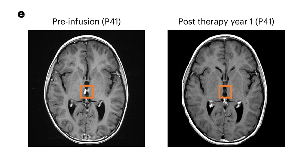
That is the trial’s most striking observation and its most fragile causal story. The tumor’s natural history is incompletely defined. The amount of viable tumor at baseline was difficult to confirm. Re-irradiation and chemotherapy came before the cells. The response was delayed, and there was no control group.
The immune evidence does not close that gap. The study used an ELISpot assay, a laboratory test that detects signals released by individual T cells when they react to a chosen target. P41’s manufactured cells tested negative under the study’s original analysis rules. A positive result appeared only under a less strict reanalysis and was later removed because of false-positive concerns; the test plate was cracked, leakage was possible and no product remained for retesting. The researchers also could not track the infused cells by their unique T-cell-receptor fingerprints, called clonotypes. No product remained to establish which fingerprints belonged to the infusion, and later mixed immune-cell samples from blood were used only in a broad analysis, not a direct product-to-blood match. The available samples therefore could not directly establish long-term persistence or expansion of cells from the infusion product.
What can these immune tests show?
ELISpot asks whether T cells release a signal when exposed to a selected target in the laboratory. Receptor-fingerprint tracking asks whether the same T cell families found in the infusion product can later be found and measured in blood or tissue. A convincing result could support a chain from target recognition to persistence, but it would still not prove that the cells caused a clinical response. Here, technical problems and missing matched samples left even that supporting chain incomplete.
The study therefore did not demonstrate that P41’s infused cells recognized the intended targets, remained in the body or caused the complete response.
P35: more than 31.8 months, then follow-up ended
P35 entered at age 14 with grade 4 medulloblastoma, a high-grade brain cancer, first diagnosed at age 7. By enrollment, the disease had returned three times and the patient had received 17 disease-directed treatments.
After TAA-T, the paper reports prolonged stability on scans without additional antitumor treatment. The source data document a disease-free interval of more than 31.8 months from the first infusion. At that point P35 left the study. In study statistics, the observation was censored: follow-up stopped there, without assuming what happened afterward. It must not be extended to the paper’s final cutoff.
At baseline, the disease could not be measured dependably or assessed well enough on scans to classify a response. The extensive treatment history and absence of a randomized comparator make it impossible to assign the interval to one intervention.
P36: surgery and chemotherapy before, targeted treatment after
P36 entered at age 14 with pediatric glioblastoma that had returned after progression on standard chemotherapy plus radiation and an investigational PARP inhibitor, a drug intended to block one route tumor cells use to repair damaged DNA. The tumor progressed about three months before the first infusion; repeat surgery and temozolomide chemotherapy followed before TAA-T.
No disease-directed therapy was given during active TAA-T treatment, but after the final infusion P36 received seven months of a CDK4/6 inhibitor, a targeted drug designed to slow cell division. The source data document a disease-free interval of more than 51.6 months from the first infusion, ongoing at the study cutoff.
That interval is the longest of the three. It also sits inside a sequence containing surgery, chemotherapy, TAA-T and later targeted treatment. The trial cannot separate their effects.
Why “after” cannot become “because of”
These vignettes matter precisely because their limits are kept beside them.
ReMIND was not randomized, had no concurrent control group and was not large enough or designed to test whether the treatment worked. Arms B and C combined different diagnoses, amounts of disease, prior treatments and expected disease courses; pooling them was a descriptive choice made after the researchers saw the data. Arm C added pre-infusion immune-lowering chemotherapy, while arm B did not. Some patients pursued additional therapy after TAA-T. The people who remained without progression had very little tumor visible at the start, which may itself influence outcome.
ClinicalTrials.gov and the paper count participation differently. The registry currently lists 33 people as enrolled and focuses its main safety measure on the first 42 days. The paper and protocol begin with 51 people who consented, report 33 who were infused, and describe their main questions as safety, whether the treatment could be made and delivered, and which dose should move forward. This article uses the paper and protocol’s definitions rather than combining the two counting systems.
None of those caveats make the three intervals unimportant. They determine what kind of importance the evidence can carry.
The result is a preliminary signal: a reason to test the strategy in a study designed to estimate benefit, with clearer biological measurements and a comparison group able to separate treatment from selection, timing and other care. It is not evidence that three children were cured by T cells. It is not evidence that the cells crossed the blood-brain barrier—the protective boundary between circulating blood and brain tissue—and killed the tumors. It is not a treatment families can currently request outside research.
What comes next
ReMIND’s practical question—could the treatment be made and delivered?—needs two counts. A TAA-T product at the trial’s first planned dose or higher was manufactured for 41 of the 48 patients whose blood was collected, while 33 of the 51 consented patients received at least one infusion. The process of making the treatment also improved during the trial. The clinical histories justify further work, but the next study must ask a different question.
Researchers will need to establish whether the cells reliably recognize their intended targets, where they travel, how long they persist and whether outcomes improve compared with other care. Larger studies will also be needed to characterize uncommon harms. A future trial designed to test benefit must preserve the distinctions this phase 1 study could not resolve: tumor type, amount of disease, pre-infusion chemotherapy, prior treatment and treatment given afterward.
For families facing pediatric brain tumors, uncertainty is not the same as emptiness. Three long intervals can be worth attention without being turned into a promise. The most respectful version of hope is the one that tells the truth about how much remains unknown.
Editorial note: what the numbers cannot carry
It is natural to wish that this story ended with every child cured. It does not. ReMIND was conducted in diseases for which families can face terrible choices and clinicians must act without knowing whether an experimental treatment will help, have no effect or cause harm.
The study recorded long disease-free intervals. It also recorded severe adverse events and the death of P27. The investigators judged that fatal event possibly related to TAA-T and probably related to the underlying disease; imaging was consistent with progression. That uncertainty cannot be resolved after the fact, and it must not be turned into blame.
The code P27 protects a child’s privacy. It should not make the child abstract. Behind every patient number was a young person, a family making decisions under pressure and a clinical team responsible for care in circumstances where no option came with certainty. Participation did not oblige any of them to produce a hopeful or “heroic” outcome, and a death does not become an acceptable price simply because later research may learn from it.
Medical progress is not made only by treatments that cure. A phase 1 study can also establish what can be manufactured and delivered, identify a dose for further testing, show which harms require attention and indicate how a protocol must change. That knowledge does not redeem suffering. It creates an obligation to report it honestly, use it to make later studies safer and remember the people who made it possible.
Clean summary
ReMIND was an early phase 1 trial of T cells grown from each participant’s own blood and expanded in the laboratory to react to three proteins that can be present in cancer cells. Of 51 children and young adults with high-risk brain or spinal-cord tumors who consented, 48 had blood collected, 41 had a product manufactured at the first planned dose or higher, and 33 received at least one infusion. Most recorded health problems were mild or moderate, but three infused patients had severe or worse events considered at least possibly related; two had possibly related serious brain- or tumor-swelling events, including one death that met the trial’s dose-limiting rule. In the aggressive brainstem-tumor group, no scan showed enough shrinkage or disappearance to meet the trial’s predefined response threshold. In the non-brainstem groups, three young patients had disease-free intervals beyond 31.8, 41.2 and 51.6 months from first infusion, including one complete response under the scan criteria. Follow-up ended when one patient left the study; the other two intervals were ongoing at the cutoff. The trial had no control group, was not designed to establish benefit, and cannot show that the experimental T-cell therapy caused those outcomes.
No-BS check
What the paper shows: A personalized T cell product aimed at three tumor-associated proteins was made at the trial’s first planned dose or higher for 41 of 48 patients whose blood was collected; 33 of 51 consented patients received at least one infusion. A statistical model selected a dose for later testing. The trial documented the health problems that occurred and recorded three long disease-free intervals after infusion.
What is promising but unproven: That the experimental T cells contributed to disease control in some patients, especially those with very little tumor visible at the start, and could become clinically useful after controlled testing.
What it does not show: That the treatment cured three children; that it caused the complete response or prolonged intervals; that P41’s infused cells were demonstrated to recognize the intended targets or remain in the body; that the approach works in this aggressive brainstem tumor; or that the treatment is available or broadly safe.
Main limitations: This was an early safety-and-dose study in which everyone knew the treatment and no one was randomly assigned to a comparison group; 33 patients with different diagnoses were infused; early signs of benefit were not the main question; survival was measured from different starting points across groups; arm C contained only four infused patients and used pre-infusion immune-lowering chemotherapy; some patients received other therapies; the three exceptional cases did not begin with a tumor area that could be measured reliably on scans—P41 still had visible disease that doctors could follow, while P35 and P36 could not be evaluated well enough to classify a response; laboratory evidence could not directly link the infused cells to the later outcomes; and one fatal event met the trial’s most severe rule for toxicity.
How much confidence should a general reader have? High confidence in the narrow findings about making and delivering the treatment and the health problems recorded in this group. High confidence that the three documented intervals occurred after infusion. Low confidence that the experimental T-cell therapy caused those outcomes, because this trial was not designed to answer that question.
Sources
Based on: Multi-antigen-targeting T cells in pediatric central nervous system tumors: a phase 1 trial — Stephanie Gomez, Rachel A. DiCioccio, Ashley E. Geiger, Melanie L. Grant, Emily Reynolds, Anushree Datar, Chase D. McCann, Jay Tanna, Divyesh Kukadiya, Fahmida Hoq, Anqing Zhang, Patrick J. Hanley, Jennifer L. Webb, Lindsay B. Kilburn, Brian R. Rood, Adriana Fonseca, Holly J. Meany, L. Gilbert Vezina, Roger J. Packer, Conrad Russell Y. Cruz, Catherine M. Bollard & Eugene I. Hwang, Nature Medicine 32, 2481-2493 (2026).
- Paper — Gomez et al., Multi-antigen-targeting T cells in pediatric central nervous system tumors: a phase 1 trial, Nature Medicine 32, 2481-2493 (2026)
- Source — Author Correction, Nature Medicine (22 July 2026), DOI 10.1038/s41591-026-04593-2
- Source — ClinicalTrials.gov, NCT03652545
The source article was followed by an Author Correction that clarified which patent applications the institution had instructed counsel to abandon. The correction did not change the trial results.
Written by
Cinzia Vaglio · figures and links by
Cinzia Vaglio, Nova Vela · edited by
Laura Nesso, Michele Renda
· figures and links by
Cinzia Vaglio, Nova Vela · edited by
Laura Nesso, Michele Renda
The Clean Paper · 2 August 2026 · Text on this site is licensed under CC BY-SA 4.0. Images are licensed separately. 


Editorial note
This article was written by AI and reviewed by the editorial team. It is a clear, conservative explanation of the linked work, not a substitute for reading it. Responsibility for selection, interpretation, and final wording rests with the editor.